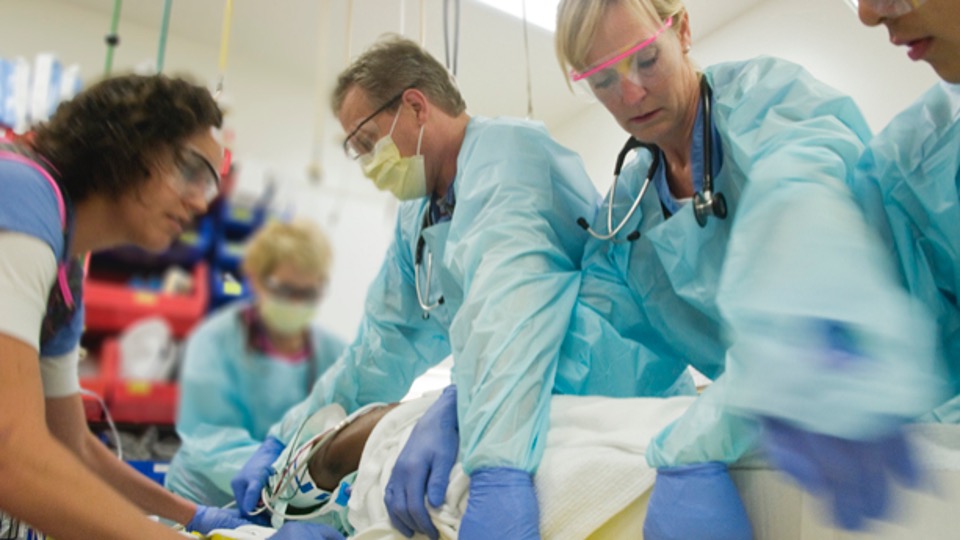
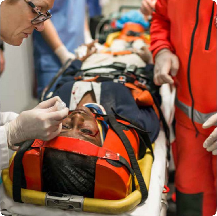
ATLS was created to standardise the first hour of trauma care — so any clinician, in any hospital, can deliver a safe, structured approach to immediately life-threatening problems.
Key timeline
February 1976: Dr James K. Styner crashed his plane in rural Nebraska. Noticing a worrying lack of emergency trauma training at the local hospital, he highlighted major variability and gaps in trauma care.
1978: The first prototype ATLS course was delivered locally, turning the "what should we do first?" problem into a teachable system.
1980: The American College of Surgeons (ACS) adopted and began wider dissemination of ATLS.
Take-home: ATLS is a shared language — prioritise threats, intervene early, reassess repeatedly.
🎯 Learning objectives
By the end of this module, you should be able to:
Explain the purpose and philosophy of ATLS.
Describe the primary survey using the ABCDE approach.
Recognise life-threatening injuries.
Understand the difference between primary and secondary survey.
Appreciate the importance of team roles and communication.
Apply ATLS principles to a trauma case.
⚠️ This module is not a substitute for the formal ATLS course delivered by the Royal College of Surgeons of England. It is designed as a brief overview and supplementary learning resource.
⚙️
ATLS · Major Trauma
Trauma Call Workflow at WSH
What happens from alert to primary survey
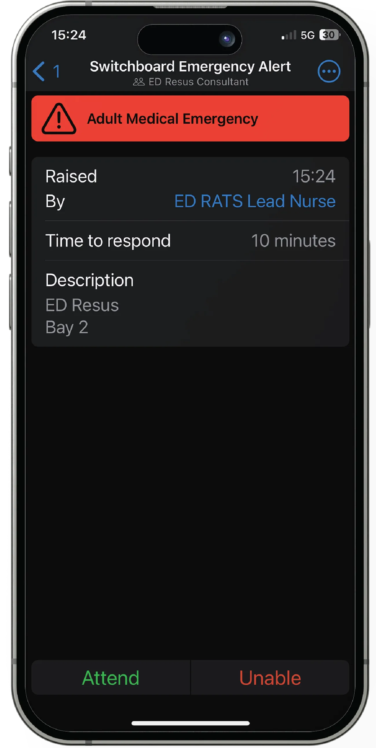
Alertive critical alert
1. Trauma Alert Activated A critical alert is sent on Alertive for adult or paediatric trauma call.
↓
2. Trauma Team Assembles Team members attend the designated resus bay, introduce themselves, and prepare for arrival.
↓
3. Sign-in Team members sign the trauma call booklet to confirm attendance.
↓
4. Patient Arrival & Handover Patient transferred to resus trolley; paramedics deliver a structured handover.
↓
5. Primary Survey Begins Trauma assessment proceeds immediately to the ABCDE primary survey.
The complete trauma assessment has 10 stages. Click each to expand:
1PreparationSet up team and environment before patient arrives.Expand ▾
Team readiness: roles allocated, PPE, resus bay prepared, airway/IV/monitoring equipment checked, trauma call activated.
2TriagePrioritise based on physiology, mechanism and severity.Expand ▾
Rapid prioritisation based on physiology, mechanism, and suspected severity to ensure the sickest patients are managed first.
3Primary SurveyIdentify and treat life-threatening problems (ABCDE).Expand ▾
Airway with C-spine, Breathing, Circulation/haemorrhage control, Disability, Exposure/temperature — identify and treat immediately life-threatening problems.
4ResuscitationTreat life threats while you assess.Expand ▾
In an MTC, the core team is supplemented by on-site specialties for definitive trauma care.
🩸 Vascular Surgery
Major vascular injury and haemorrhage control.
❤️ Cardiothoracic Surgery
Major thoracic/cardiac injury, complex chest trauma.
✋ Plastic Surgery
Soft tissue injury, limb salvage, complex wounds.
🧠 Neurosurgery
Severe TBI, intracranial injury, spinal injury.
ABCDE
ATLS · Quick Reference
Primary Survey (ABCDE)
Identify and treat immediately life-threatening problems. Treat as you find. Re-check after every intervention.
Video
Primary Survey Demonstration
Demonstration of a structured ATLS primary survey (ABCDE approach)
Tap to expand each step:
AAirway with C-spine controlIs the airway patent? Protect the neck.Expand ▾
Goal: Prevent hypoxia and secondary spinal cord injury.
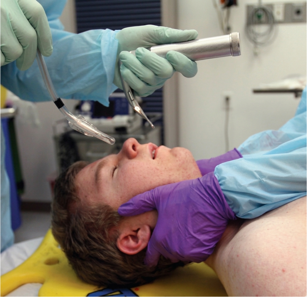
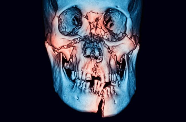
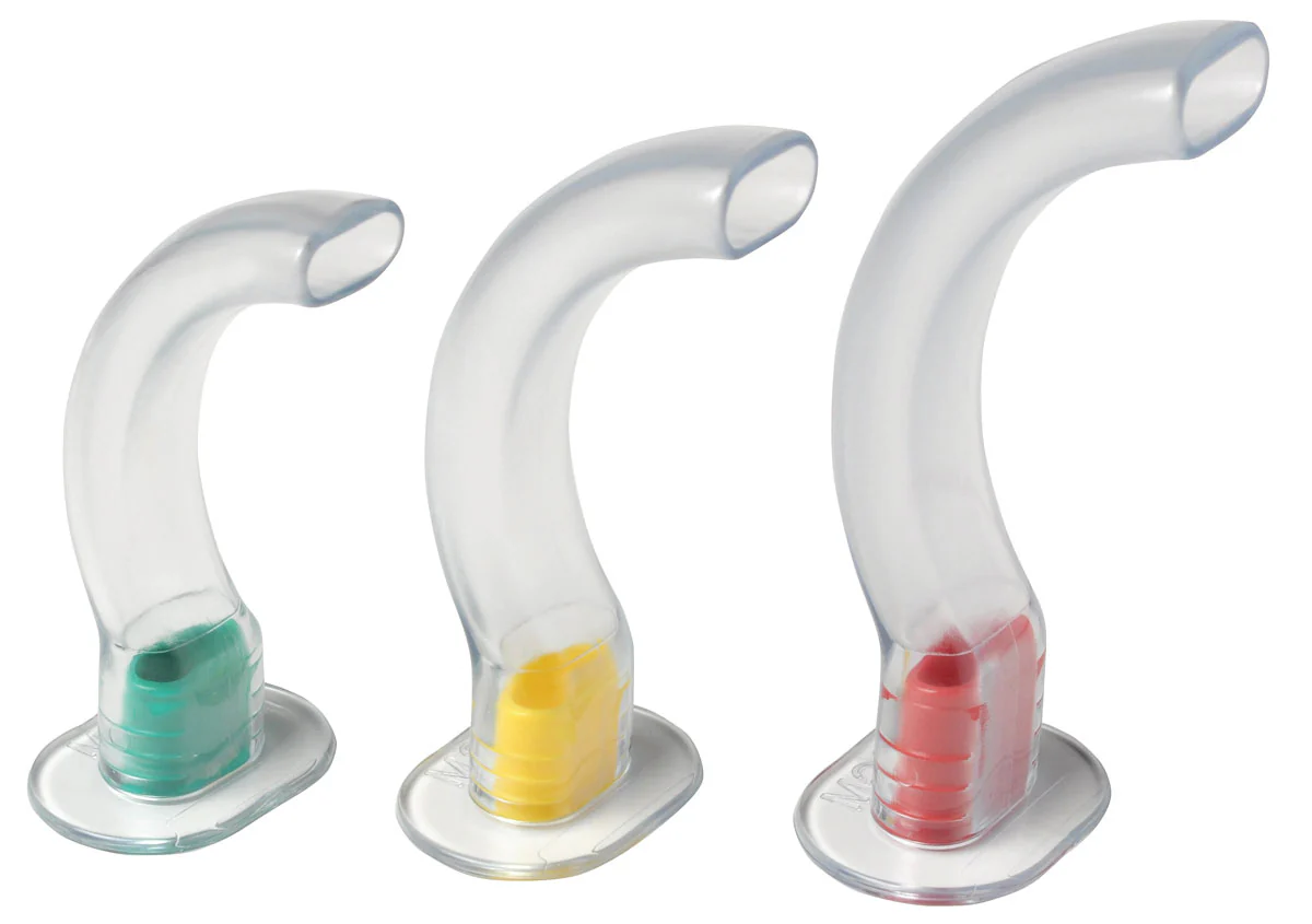
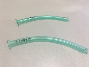
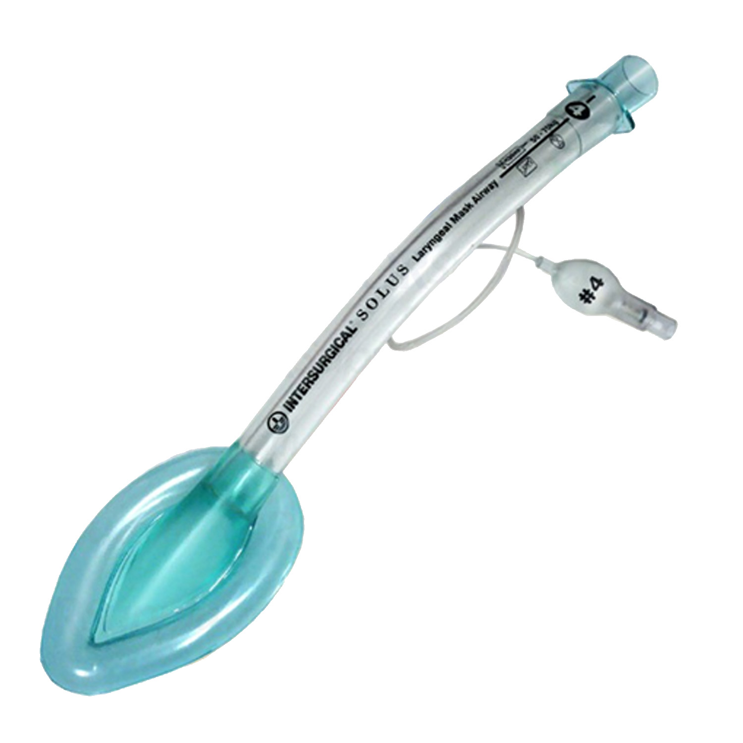
Assess: Speech, airway sounds (snoring/stridor/gurgling), facial trauma, vomit/blood, burns, swelling. Act: Jaw thrust + suction, high-flow O₂, airway adjuncts (OPA/NPA if appropriate), manual in-line stabilisation, early anaesthetic input if concern.
BBreathing & VentilationIs oxygenation and ventilation adequate?Expand ▾
Goal: Prevent hypoxia and ventilatory failure.
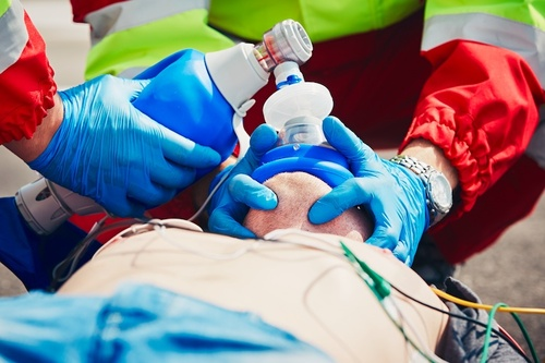
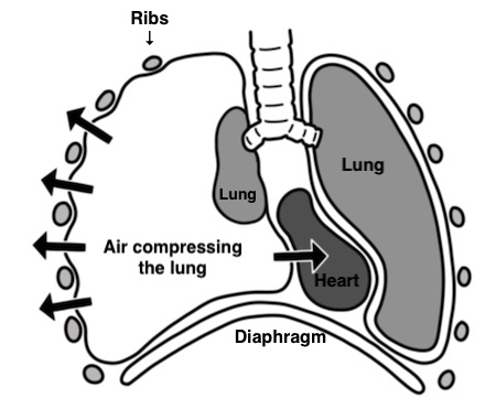
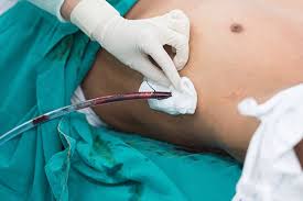
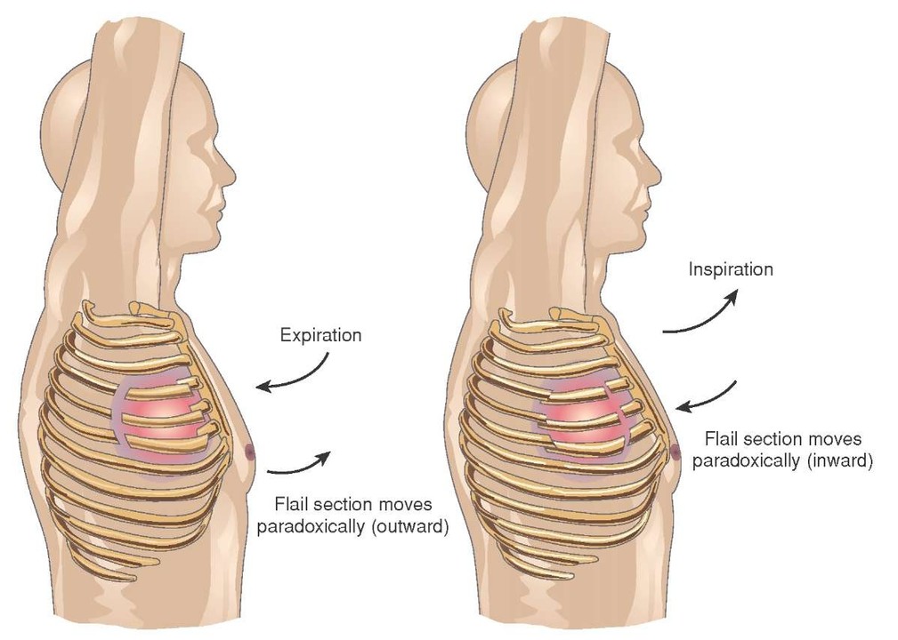
Assess: RR, SpO₂, work of breathing, chest expansion, trachea, breath sounds, wounds/bruising. Act: Oxygen, ventilatory support if needed, seal open pneumothorax, decompress suspected tension pneumothorax, analgesia for rib fractures.
CCirculation with Haemorrhage ControlStop bleeding first, then restore perfusion.Expand ▾
Goal: Prevent death from haemorrhagic shock.
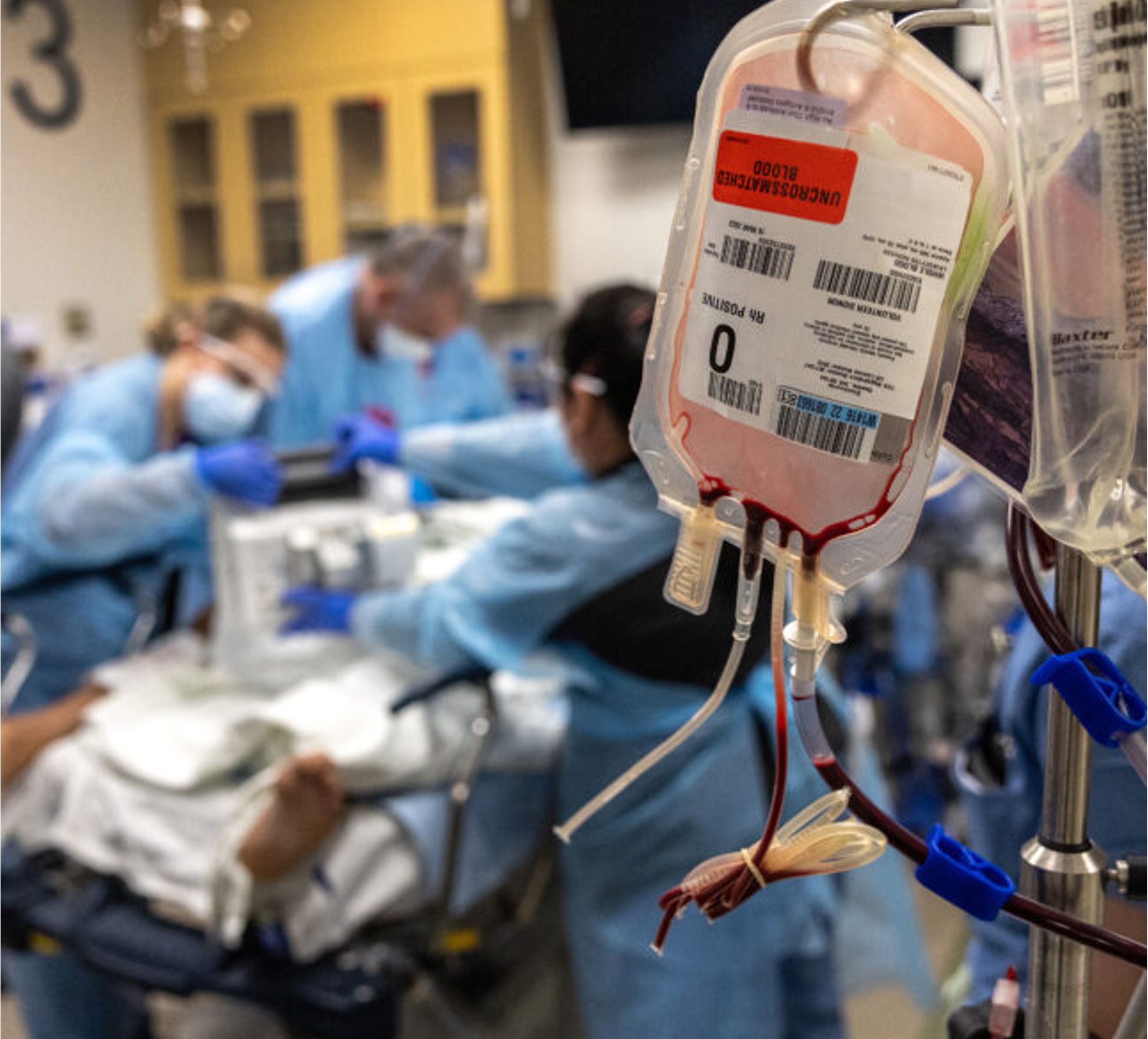
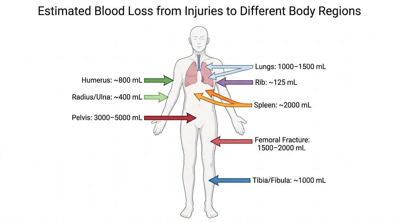
Assess: Major external bleeding, pulse, BP, cap refill, skin temperature, likely internal sources (chest/abdomen/pelvis/long bones). Act: Direct pressure/haemostatic dressing, tourniquet if indicated, pelvic binder if suspected, IV/IO access, bloods + group & save, activate major haemorrhage protocol early.
Assess: Fully expose (front/back when safe), look for hidden bleeding, deformity, penetrating injury. Act: Warm blankets, warmed fluids, minimise exposure time, document temperature early.
Key principle: ABCDE is done in order. Do not move on if a life-threatening issue is identified. Intervene immediately and re-assess from the top.
A
ATLS · Primary Survey
Airway with C-spine Control
Airway first — obstruction → hypoxia → death within minutes. Manage while protecting the cervical spine.
Fast check: Talking in full sentences = airway patent. Stridor, gurgling, or silence = airway compromise.
C-spine rule: In trauma, assume cervical spine injury until proven otherwise. Airway assessment and C-spine protection should occur simultaneously by different team members.
Tip: document both positives and key negatives to improve handover and reduce missed injuries.
65+
ATLS · Major Trauma
Silver Trauma
Serious injury with apparently minor mechanisms. Physiological reserve is reduced — escalate earlier.
Think early about (any patient 65+)
Mechanism that may be underestimated
Physiological abnormality even if subtle
Anticoagulation / coagulopathy
Head, chest, pelvic, and spinal injury
Frailty, comorbidity, and baseline function
Infographic
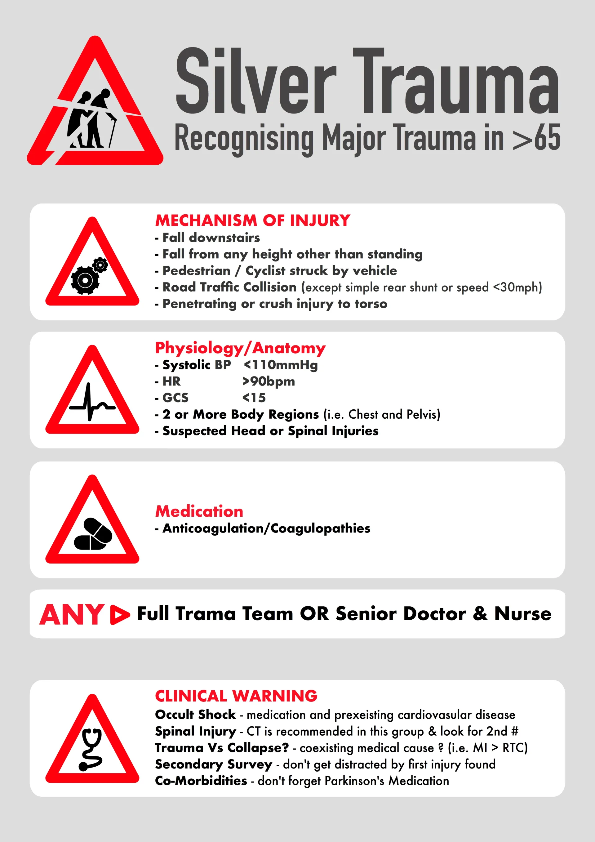
Silver trauma triggers and warning signs
Key concept
Occult shock and under-triage
Older patients may not mount the expected tachycardic or hypotensive responses due to beta-blockers, pacemakers, or limited physiological reserve.
SBP <110 may already indicate significant compromise
GCS <15 in an older trauma patient is concerning
Low-energy mechanisms can still produce major injury
Do not miss
Occult shock
Intracranial bleeding, especially on anticoagulation
Spinal injury after apparently minor falls
A second injury found only on secondary survey
Important regular medications e.g. Parkinson's medication
ICH
ATLS · Neurosurgical Trauma
Intracranial Haemorrhage in Trauma
Haemorrhage types, key actions after CT, and neurosurgical referral pathway.
Core message
ICH can deteriorate quickly. Priorities after CT: prevent secondary brain injury, reverse anticoagulation where relevant, and obtain a documented neurosurgical plan.
Red flags — escalate if:
Falling GCS, new focal deficit, seizures
Abnormal pupils / signs of herniation
Anticoagulated patient with any bleed
Significant midline shift on CT
Types of intracranial haemorrhage — Learn by clicking each type
Quick check
Test your knowledge
Q1: A patient with a temporal fracture develops a "lucid interval" then deteriorates rapidly. Which type of bleed?
A) Epidural Haematoma
B) Subdural Haematoma
C) Subarachnoid Haemorrhage
D) Parenchymal Haemorrhage
Q2: An 85-year-old on warfarin has a subdural bleed. Which is the correct shape?
A) Crescent (follows dural folds)
B) Lens-shaped (convex)
C) Diffuse in sulci
D) Focal mass in brain
Key actions after CT confirms a bleed
Prevent secondary brain injury: avoid hypoxia and hypotension; correct glucose and temperature.
Clarify anticoagulation/antiplatelets: last dose time, indication; send coagulation bloods.
Urgent neurosurgical referral by A&E prior to T&O referral — request a clear written plan.
Document escalation triggers: fall in GCS, pupillary change, seizures, worsening headache/vomiting.
⚠️ Do not accept the patient under T&O until there is a documented plan from neurosurgery confirming the patient is for local/conservative management.
Anticoagulants — Reversal (Practical Summary)
In suspected or confirmed intracranial haemorrhage on anticoagulants, A&E should discuss reversal urgently with the on-call haematologist (and ensure neurosurgery are aware) and document the agreed plan.
What to prepare before you call
Drug(s) + time of last dose (warfarin / DOAC / antiplatelet)
Indication for anticoagulation (AF, VTE, valve, etc.)
Renal function (U&E), coagulation profile (INR/APTT), FBC
CT summary + current neuro status trend
What you should record
Name/grade of haematologist and time of discussion
Reversal plan and monitoring (e.g. repeat INR/coags)
Prescribe the reversal agent (e.g Octaplex prescribed on pink transfusion prescription form)
☐ GCS documented (trend) ☐ Pupils documented ☐ Glucose checked
☐ CT head reviewed: type of bleed recorded
☐ Anticoagulant/antiplatelet status clarified
☐ Haematology discussed if anticoagulated: plan documented and actioned
☐ Neurosurgery contacted and plan recorded in notes
☐ Neuro obs frequency + repeat CT timing + escalation triggers documented
🦴
ATLS · Pelvic Trauma
Pelvic & Acetabular Injuries
Recognise instability, control haemorrhage early, identify associated injuries, and escalate promptly.
Core message
Pelvic trauma can cause life-threatening haemorrhage. Early priorities: treat haemorrhage, stabilise the pelvis, and escalate to the right team.
Think pelvic bleeding if:
High-energy mechanism + pelvic pain/deformity
Haemodynamic instability without another clear source
Unstable pelvic ring injury on imaging
Ongoing transfusion requirement
Pelvic Ring Injuries — Interactive Activity
Click each injury type to explore its mechanism, stability, and haemorrhage risk. Then test your knowledge.
Teaching point: The more disrupted the posterior ring, the more unstable and dangerous the injury. Vertical shear injuries carry the highest risk of life-threatening haemorrhage.
Once you've reviewed all four types, test your knowledge below.
Q1. A motorcyclist is hit from the side. Imaging shows bilateral pubic rami fractures and a sacral compression fracture. Which pelvic ring injury pattern is this?
AAPC — Anteroposterior Compression
BLC — Lateral Compression
CVS — Vertical Shear
DCM — Combined Mechanism
Q2. A 32-year-old falls two storeys from scaffolding. AP pelvis X-ray shows superior displacement of the right hemipelvis. What is the single most important reason this fracture pattern is dangerous?
AIt causes severe pain that limits assessment
BDisrupted posterior ligaments cause haemodynamic instability from massive haemorrhage
CThe sciatic nerve is directly lacerated by bone fragments
DHigh risk of bowel perforation
Q3. Which pelvic ring injury pattern increases pelvic volume and therefore poses the greatest early risk of retroperitoneal haemorrhage?
AAPC — Open-book injury
BLC — Lateral Compression
CVS — Vertical Shear
DCM — Combined Mechanism
Immediate Management
☐ Escalate to a senior SpR or Consultant if not already involved
☐ Treat haemodynamic instability as haemorrhage until proven otherwise
☐ Minimise pelvic movement
☐ Apply pelvic binder early if suspected unstable pelvic ring injury over greater trochanters
☐ Activate major haemorrhage protocol if unstable and suspected pelvic bleeding
☐ Screen for associated injuries: GU, bowel, spine, long bones
☐ Imaging: pelvic X-ray early if unstable; CT when stable enough
☐ VTE prophylaxis plan documented:
If surgery/transfer anticipated → anticoagulants may be held if safe.
If urgent transfer is not required and haemostasis is secure → start VTE prophylaxis
Referral Workflow
Urgent discussion via the Major Trauma Network if: (to be arranged by A&E)
Haemodynamic instability with suspected pelvic bleeding
Unstable pelvic ring injury / ongoing transfusion requirement
Open pelvic fracture, vascular injury, significant GU injury
Complex acetabular fracture, hip dislocation with fracture, neurovascular deficit
For stable but complex pelvic and acetabular injuries
Our local pelvic and acetabular surgical team are based in Addenbrookes Hospital
Transfer any relevant images to Addenbrookes Hospital
Select New referral → Addenbrookes Hospital →
Complex Orthopaedic Trauma and fill in the referral form
Isolated Pubic Rami Fractures
If there is no concern of further pelvic injury these patients can be conservatively managed as a fragility fracture with analgesia and physiotherapy
Refer to the medical team as per the trust's Emergency Admissions Policy
No orthopaedic follow up is required
🦴
ATLS · Spinal Injuries
Spinal Injuries
Maintain spinal precautions until injury excluded — escalate any neurological deficit urgently.
Core message
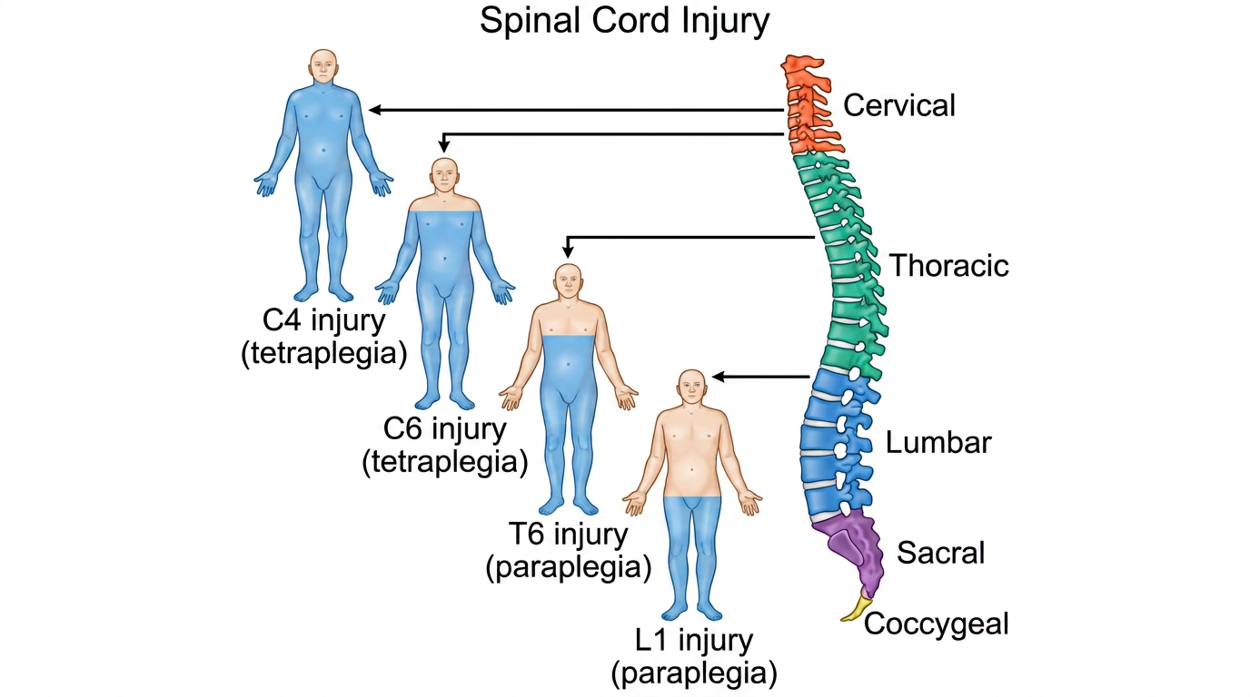
Treat any suspected spinal injury with spinal precautions until cleared. Early priorities: protect the cord, recognise neurological deficit, obtain appropriate imaging, and determine whether the injury is stable or unstable.
Escalate urgently if
Any new or evolving neurological deficit
Suspected spinal cord compression
Unstable injury pattern on CT/MRI
Persistent severe midline spinal pain/tenderness with high-risk mechanism
Types of spinal fractures — Learn by clicking each type
Question 1: Which spinal injury is most commonly associated with a seatbelt mechanism?
A) Burst fracture
B) Odontoid fracture
C) Compression fracture
D) Chance fracture
Question 2: A cervical facet dislocation is highly unstable. What is the primary risk?
A) Vertebral body collapse
B) Spinal cord injury from malalignment
C) Loss of disc height
D) Posterior ligament disruption only
Determining stability
Neurology: any deficit implies high concern and urgent escalation
Multiple levels: non-contiguous injuries — image the whole spine as appropriate
Initial management
☐ For unstable fractures or neurological deficit, escalate to SpR or Consultant
☐ Maintain spinal precautions until injury confirmed stable (collar, logroll, bed rest if required)
☐ Document neuro exam: power/sensation in limbs, bladder/bowel symptoms
☐ Analgesia
☐ Look for associated injuries (head, chest, pelvis, long bones)
☐ Catheterise
Referral to the spinal unit
Urgent referral required for:
Any neurological deficit (new or worsening)
Suspected cord compression / cauda equina features
Transfer images via PACS (9am–5pm) or on-call radiographer (5pm–9am)
Online referral: referapatient.org → New Referral → Ipswich Hospital → Spinal Surgery
8am–8pm: phone call to spinal fellow at Ipswich (usually T&O SpR)
Urgent overnight: On-Call T&O SpR at Ipswich via switchboard
📋
ATLS · Clinical Scenario 1
Mind the Cat
Trauma Assessment Scenario
Scenario
You are called to the ED
You are the orthopaedic SHO on-call. A 76-year-old woman is brought to the Emergency Department.
She recalls getting up in the night and tripping over her cat at home, but cannot remember what happened after this.
She was later found wandering around her kitchen. The ambulance crew noted blood on the corner of a side table at the foot of the stairs.
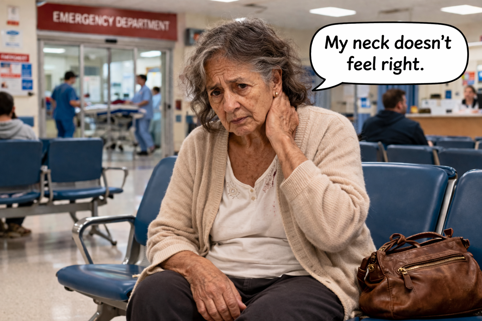
She complains of pain and tenderness at the back of her head, where there is a bleeding laceration. You notice she is holding her neck and appears uncomfortable. She looks at you and says:
"It feels unsteady."
What are your immediate priorities in the first 60 seconds?
Think through your ABCDE approach before revealing the answer.
Immediate priorities
A — Maintain manual in-line C-spine immobilisation immediately. She has a neck complaint, back-of-head injury, and possible fall mechanism — assume cervical spine injury until excluded. Do not allow her to move her neck.
B — Ensure she is breathing adequately. Apply high-flow oxygen via a non-rebreather mask. Check RR and SpO₂.
C — Assess haemodynamic status. Check pulse, BP, cap refill. The head laceration may be bleeding — assess and apply direct pressure if actively bleeding.
D — Rapid neuro screen. GCS (this is concerning — she has had LOC and cannot recall events). Check pupils, blood glucose, and limb movement.
E — Do not expose fully in the corridor, but note she is a 76-year-old (silver trauma): low threshold for CT head and CT cervical spine, and proactive temperature management.
⚠️ Silver trauma alert: Her medications (anticoagulants?), antiplatelet use, and baseline frailty are critical to establish early. A seemingly minor fall in a 76-year-old can result in significant intracranial or spinal injury.
Cervical spine injury — she is holding her neck and reports instability. Maintain C-spine immobilisation and request CT cervical spine.
Intracranial haemorrhage — LOC + head laceration in an elderly patient = CT head urgently, especially if anticoagulated. Can be EDH, SDH, SAH, or contusion.
Occult shock — in a 76-year-old, normal observations do not exclude significant bleeding. A "normal" BP in an elderly hypertensive patient may still represent significant compromise.
Syncope as the primary event — consider: did she fall because she collapsed? Is there a cardiac or neurological cause for the collapse? "Trauma causing collapse" vs "collapse causing trauma".
⚠️ Pause and decide: How would you escalate this patient if her GCS dropped from 14 to 12 while you were assessing her?
📋
ATLS · Clinical Scenario 2
Break Leg Speed
Trauma Assessment Scenario
Scenario
You are called to the resus bay
You are the orthopaedic SHO on-call. A 35-year-old motorcyclist is brought in following an RTC at approximately 60 mph.
He was unable to mobilise at the scene. He reports pain in his left shoulder and severe pain in his left thigh, which appears deformed. The limb was splinted pre-hospital.
During transfer, observations deteriorated: BP 80/61, HR 120. A pelvic binder was applied en route.
As you approach, he looks distressed and says:
"My leg hurts... and I feel really dizzy."
What are your immediate priorities in the first 60 seconds?
He is shocked (BP 80/61, HR 120) — work through your ABCDE before revealing.
Immediate priorities
A — Airway with C-spine control. High-energy mechanism at 60 mph = assume cervical spine injury. He is talking (airway currently patent) but maintain in-line stabilisation.
B — Breathing. Apply high-flow O₂, check RR and SpO₂, examine the chest — is there a pneumothorax or haemothorax contributing to deterioration?
C — Circulation: this patient is in Class III haemorrhagic shock. BP 80/61, HR 120 = significant haemorrhage. Establish two large-bore IVs immediately, take bloods (FBC, U&Es, LFTs, clotting, crossmatch), activate the Major Haemorrhage Protocol, give TXA, and request blood products. The pelvic binder is already in situ — do not remove it.
D — Disability. GCS? He is communicating — but dizziness and hypotension may reduce conscious level. Check glucose.
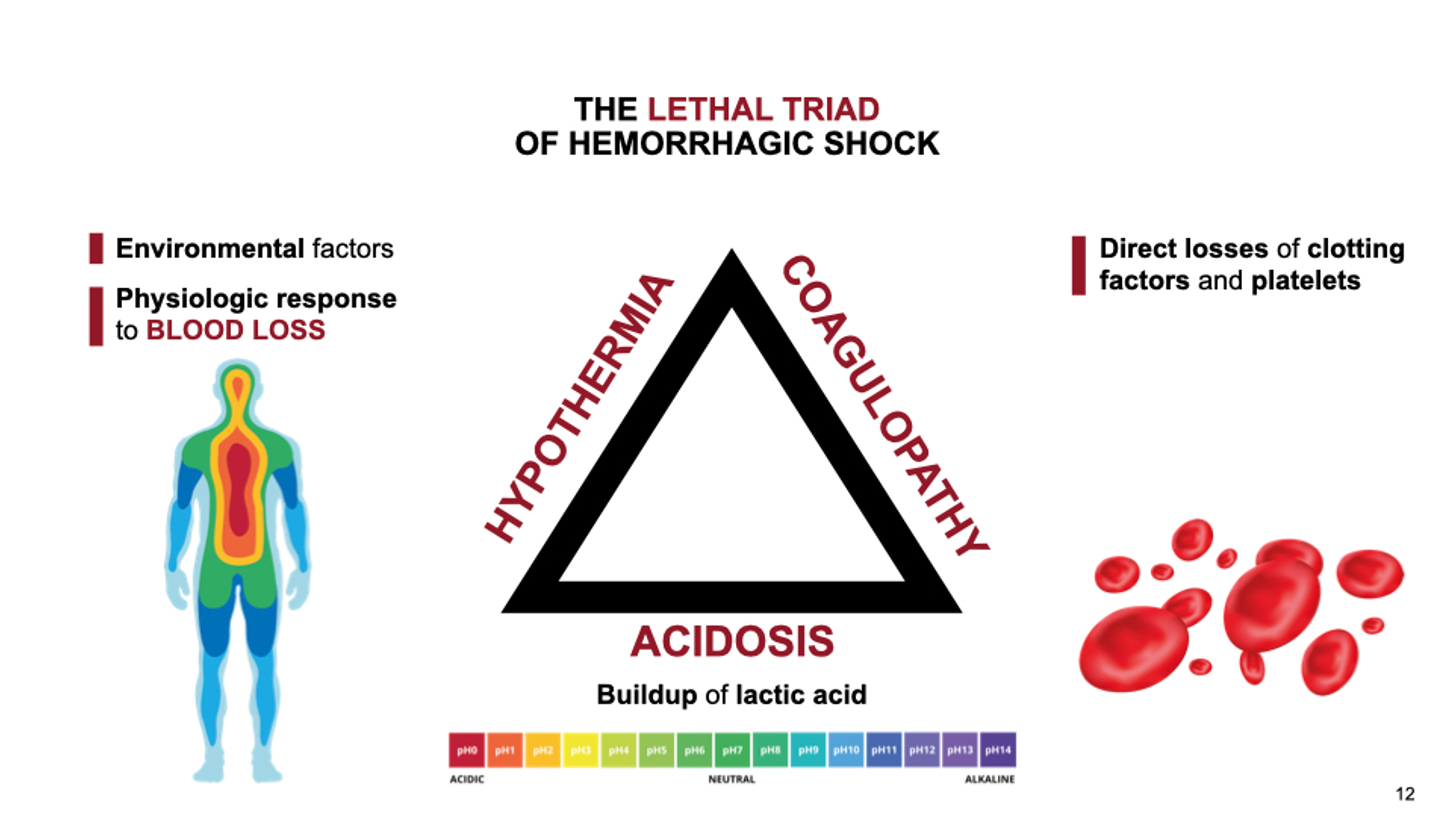
E — Exposure. Expose carefully to look for hidden injuries. Keep warm — he is at risk of the lethal triad.
What are the potential sources of life-threatening haemorrhage?
He has a femoral fracture, a pelvic binder in situ, and left shoulder pain — think "on the floor and four more".
Potential haemorrhage sources
Left femoral fracture — can lose 1500–2000 mL. Pre-hospital splinting will have reduced ongoing bleeding. Reassess the limb — is the splint secure?
Pelvis — pelvic binder suggests concern for pelvic injury. An unstable pelvic ring can bleed 3000–5000 mL. The binder reduces pelvic volume — do not remove without senior guidance.
Chest — high-energy mechanism: haemothorax or tension pneumothorax? Examine the chest formally during primary survey.
Abdomen — solid organ injury (spleen, liver) in high-energy RTC. May need FAST scan or urgent CT.
Left shoulder pain — could indicate clavicle fracture (lower haemorrhage risk) but also consider subclavian vascular injury or associated thoracic injury.
💡 Call for help now: This patient is haemodynamically unstable. Your SpR, the ED consultant, anaesthetics, and the on-call radiologist (for potential IR) should all be alerted immediately.
⚠️ Pause and decide: After your primary survey, his BP remains 70/50 despite 2 units of blood. What do you do next?
🧠
ATLS · Knowledge Check
Knowledge Check
Test your understanding of the key principles from this module.
Question 1 of 5
A trauma patient is talking but making gurgling sounds. Which ABCDE step takes priority and what is the most important immediate action?
AB — Breathing: apply high-flow O₂ via non-rebreather mask.
BD — Disability: check GCS and pupils immediately.
CA — Airway: jaw thrust with manual in-line stabilisation and suction to clear secretions.
DC — Circulation: establish IV access and take bloods.
Question 2 of 5
A 45-year-old following a high-speed RTC has HR 130, BP 85/50, cool peripheries, and a deformed right femur. What class of haemorrhagic shock is this, and what volume of blood may a femoral fracture lose?
AClass II shock; femoral fracture loses approximately 500–800 mL.
BClass III shock; femoral fracture can lose 1500–2000 mL.
CClass IV shock; femoral fracture can lose up to 5000 mL.
DClass I shock; femoral fracture loses less than 500 mL.
Question 3 of 5
You suspect a tension pneumothorax in a trauma patient. The patient is hypoxic and hypotensive with reduced breath sounds on the left and tracheal deviation to the right. What is the correct immediate management?
ARequest urgent portable CXR to confirm before any intervention.
BInsert a chest drain in the left 5th intercostal space immediately.
CThis is a clinical diagnosis — perform immediate needle decompression without waiting for imaging.
DIncrease oxygen flow and repeat vital signs in 5 minutes before intervening.
Question 4 of 5
An 80-year-old on warfarin trips on the stairs and presents with a GCS of 14, headache, and vomiting. CT head shows a subdural haematoma. What should you NOT do before accepting this patient under Trauma & Orthopaedics?
ADocument GCS trend and pupillary responses.
BDiscuss anticoagulant reversal urgently with haematology.
CAccept the patient under T&O without first obtaining a documented neurosurgical management plan.
DCheck the patient's INR and coagulation profile.
Question 5 of 5
Which of the following best describes the "lethal triad" in major trauma, and why is hypothermia particularly dangerous?
AHypoxia, hypotension, and hyperglycaemia — hyperglycaemia impairs wound healing.
BTachycardia, hypovolaemia, and hypoxia — all three directly reduce cardiac output.
CHypothermia, acidosis, and coagulopathy — they are self-reinforcing: hypothermia worsens coagulopathy which worsens bleeding, which worsens hypothermia.
DHypothermia, hypertension, and bradycardia — together these indicate neurogenic shock.
0/5
🎓
ATLS / Major Trauma
Module Complete
Well done for completing the ATLS / Major Trauma online module.
🏅
Congratulations!
You have completed the ATLS / Major Trauma e-learning module. This module is part of the Orthopaedic Emergencies Clinical Education Series.
What you have covered
Module Summary
📖 Introduction
Origins of ATLS, trauma call workflow at WSH, and the trauma team.
🔤 Primary Survey
ABCDE approach — Airway, Breathing, Circulation, Disability, and Exposure.
📋 Secondary Survey
Head-to-toe assessment, AMPLE history, and documentation.
65+ Silver Trauma
Physiological reserve, occult shock, and common pitfalls in older patients.
🧠 Intracranial Haemorrhage
Types of ICH, anticoagulant reversal, and neurosurgical referral pathway.
🦴 Pelvic & Spinal Trauma
Pelvic ring injury patterns, spinal fractures, and management escalation.
📋 Clinical Scenarios
Applied ATLS assessment in two realistic trauma cases.
🧠 Knowledge Check
Five-question quiz covering key principles from across the module.
⚠️ Important reminder: This module is not a substitute for the formal ATLS course delivered by the Royal College of Surgeons of England. It is intended as supplementary e-learning. For formal certification, please attend an accredited ATLS course.
Next steps
Consolidate your learning
Review any sections where you felt less confident using the sidebar navigation.
Revisit the Knowledge Check (page 18) if you wish to test yourself again.
Attend a formal ATLS course through the Royal College of Surgeons of England.
Consider completing the other modules in the Orthopaedic Emergencies series.
Apply these principles during your clinical practice — refer back as needed.
📚
ATLS / Major Trauma
References & Further Reading
Key references, guidelines, and resources underpinning this module.
Core Text
ATLS — Advanced Trauma Life Support
American College of Surgeons Committee on Trauma.Advanced Trauma Life Support (ATLS) Student Course Manual. 10th ed. Chicago: American College of Surgeons; 2018.
NICE.Head injury: assessment and early management (NG232). London: National Institute for Health and Care Excellence; 2023. Available at: nice.org.uk/guidance/ng232
NICE.Spinal injury: assessment and initial management (NG41). London: NICE; 2016 (updated 2022). Available at: nice.org.uk/guidance/ng41
NICE.Major trauma: assessment and initial management (NG39). London: NICE; 2016 (updated 2022). Available at: nice.org.uk/guidance/ng39
Haemorrhage & Resuscitation
Key papers & protocols
CRASH-2 Trial Collaborators. Effects of tranexamic acid on death, vascular occlusive events, and blood transfusion in trauma patients with significant haemorrhage. Lancet. 2010;376(9734):23–32.
Spahn DR, Bouillon B, Cerny V, et al. The European guideline on management of major bleeding and coagulopathy following trauma (STOP THE BLEED). Crit Care. 2019;23(1):98.
National Blood Transfusion Committee. Patient Blood Management Guidelines. transfusionguidelines.org
Neurotrauma
Intracranial haemorrhage & TBI
Stocchetti N, Maas AIR. Traumatic intracranial hypertension. N Engl J Med. 2014;370(22):2121–30.
Brain Trauma Foundation.Guidelines for the Management of Severe Traumatic Brain Injury. 4th ed. 2016. Available at: braintrauma.org
NHS England. Anticoagulation reversal in acute intracranial haemorrhage: NICE pathway and local formulary guidelines. (Reference local trust policy for specific agents.)
Pelvic & Spinal Trauma
Classification & management
Young JWR, Burgess AR. Radiologic Management of Acetabular Fractures. Baltimore: Urban & Schwarzenberg; 1987. (Original APC/LC/VS pelvic ring classification.)
Tile M. Pelvic ring fractures: should they be fixed? J Bone Joint Surg Br. 1988;70(1):1–12.
Trauma Audit and Research Network (TARN) — National trauma outcomes data: tarn.ac.uk
SIGN Guideline 110.Early management of patients with a head injury. Edinburgh: Scottish Intercollegiate Guidelines Network; 2009 (updated 2023).
Orthopaedic Trauma Association (OTA) — Classification and educational resources: ota.org
Disclaimer: This module is intended as supplementary educational content for healthcare professionals and does not constitute clinical guidance. Clinical decisions should always be based on current guidelines, local trust protocols, and individual clinical judgement. Content is reviewed periodically but may not reflect the most recent updates to all referenced guidelines. Always refer to your local trust protocols for patient-specific management.