Septic Arthritis
Learning Objectives
By the end of this module, you should be able to confidently recognise, investigate, and initiate management for patients with suspected septic arthritis.
Septic Arthritis
Why This Matters
Septic arthritis is an important orthopaedic emergency. A delay in diagnosis can lead to rapid cartilage destruction, permanent joint damage, systemic sepsis, and even death.
Because early symptoms can resemble other conditions such as gout, inflammatory arthritis, or traumatic effusions, the diagnosis is sometimes missed or delayed. Clinicians must therefore maintain a high index of suspicion.
Septic Arthritis
Pathophysiology
Septic arthritis occurs when microorganisms enter the joint space and trigger a rapid inflammatory response. Because articular cartilage has limited ability to regenerate, infection can quickly lead to irreversible joint damage.
Understanding the mechanisms by which bacteria reach the joint helps clinicians recognise high-risk patients and initiate prompt investigation and treatment.

• Haematogenous spread
• Direct inoculation (surgery or trauma)
• Spread from adjacent osteomyelitis or soft tissue infection
Click each route of infection to learn more
Haematogenous Spread (Most Common)
Common sources include:
- Skin infections
- Urinary tract infections
- Respiratory infections
- Intravenous lines or bacteraemia
Direct Inoculation
This may happen following:
- Joint aspiration
- Intra-articular injections
- Arthroscopy or open surgery
- Penetrating trauma
Contiguous Spread
Examples include:
- Adjacent osteomyelitis
- Soft tissue abscess
- Septic bursitis
- Deep wound infection
Septic Arthritis
Who Is At Risk?
Although septic arthritis can occur in any patient, certain groups have a significantly higher risk of joint infection. Identifying these risk factors helps clinicians maintain a higher index of suspicion.
Patients with prosthetic joints are particularly important to recognise, as infections involving joint replacements can lead to severe complications and often require surgical management.
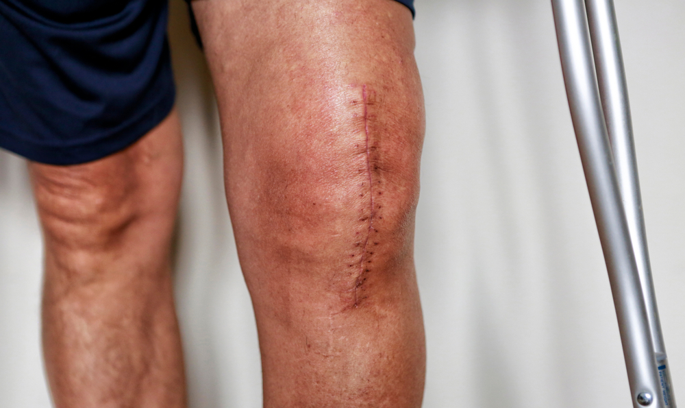
Septic Arthritis
Clinical Presentation
Patients with septic arthritis typically present with a painful swollen joint and reduced ability to move the joint. Symptoms may develop rapidly over hours to days.
The presentation can vary depending on the patient's age, immune status, and underlying joint disease. Clinicians must therefore maintain a high index of suspicion.
Examination
Examination should be focused, systematic, and gentle. Compare with the opposite side where possible, and avoid repeatedly manipulating a very painful joint.
Watch the video below for a demonstration of how to examine for fluid within the knee.
- Inspect for swelling or a visible effusion
- Look for erythema over the knee
- Assess resting position
- Check for scars from previous surgery
- Observe reluctance to move
- Compare temperature with opposite knee
- Palpate gently for tenderness
- Assess for effusion
- Identify focal tenderness or cellulitis
- Consider if joint feels tense
- Assess active range of movement
- Assess passive movement carefully
- Note if small movements cause marked pain
- Check straight leg raise or weight bearing
- Painful passive movement is a red flag
Septic Arthritis
Investigations
When septic arthritis is suspected, blood tests and imaging may support the diagnosis, but the key diagnostic investigation is joint aspiration.
- Temperature
- Heart rate
- Blood pressure
- Respiratory rate
- Oxygen saturation
Fever and tachycardia may suggest systemic infection, but normal observations do not exclude septic arthritis.
- Full blood count
- CRP
- ESR
- Urea and electrolytes
- Blood cultures
Inflammatory markers are often raised but are not specific for septic arthritis.
- X-ray of the affected joint
- Ultrasound to detect effusion
- MRI if diagnosis uncertain
Imaging may demonstrate an effusion or alternative pathology, but cannot exclude septic arthritis.
Imaging in Suspected Septic Arthritis

X-ray of the knee showing a knee effusion, marked by black arrows. It displaces the patella anteriorly and extends into the suprapatellar bursa.
- Soft tissue fullness suggesting an effusion
- Presence of prosthesis or metalwork
- Degenerative change or osteoarthritis
- Chondrocalcinosis suggesting crystal arthropathy
- Fracture, dislocation, or other structural abnormality
Septic Arthritis
Joint Aspiration
Joint aspiration is the gold standard investigation in suspected septic arthritis. It should be performed urgently to obtain fluid for microbiological analysis and crystal assessment.
If the patient is clinically stable, aspiration should ideally be performed before antibiotics are started to maximise diagnostic yield.
Watch the video below for an example of joint aspiration technique.
Prepare everything before starting the procedure so that samples can be collected and sent without delay.
- Sterile gloves
- ANTT wound pack
- Chlorhexidine spray or prep sticks
- Sterile gauze
- 10–20ml syringes
- White or green needles
- Local anaesthetic if required
- Dressing (mepilex border)
- Sharps bin
- Blood culture bottles
- EDTA bottle (red top)
- Sterile universal containers (white top)
- Request forms / labels
Once synovial fluid is obtained, it should be labelled clearly and sent promptly. Follow the flowchart below — start by selecting whether the aspiration was performed in or out of hours.
'Sterile fluid in blood culture bottle' → culture bottles
the aspiration?
9am – 5pm Out of hours
5pm – 9am
No further steps required — the lab will process directly
Await results
Note: you will need 2 universal white top containers (instead of 1)
Leave in a blue box in the biochemistry lab, or hand to the lab in the morning
Retain one at WSH · Await results
Septic Arthritis
Interpreting Synovial Fluid
Aspiration findings should always be interpreted alongside the clinical picture, observations, blood tests, and imaging.
Synovial fluid interpretation
| Condition | Appearance | WCC (cells/µL) | PMN % | Crystals | Bacteria |
|---|---|---|---|---|---|
| Non-inflammatory | |||||
| Osteoarthritis | Clear / yellow | < 1,000 | < 25% | None | None |
| Traumatic arthritis | Straw / slightly cloudy | < 2,000 | < 25% | None | None |
| Inflammatory | |||||
| Pseudogout | Cloudy yellow | 10,000–20,000 | ~70% | Calcium pyrophosphate | None |
| Gout | Cloudy / milky | 10,000–50,000 | ~70% | Urate | None |
| Rheumatoid arthritis | Cloudy yellow | 10,000–50,000 | ~70% | None | None |
| Infection | |||||
| Septic arthritis | Turbid / purulent | > 50,000 (often >100,000) | > 90% | None | Present |
Synovial fluid appearance
The image below shows synovial fluid from different conditions. Click each appearance type below to learn more about what it indicates.

Normal synovial fluid is clear and colourless or pale straw, with high viscosity. It should look similar to egg white.
- WCC < 200 cells/µL
- PMN < 25%
- No crystals, no bacteria
Straw-coloured, transparent fluid with good viscosity is typical of non-inflammatory conditions. The fluid is clear enough to read newsprint through it.
- WCC < 2,000 cells/µL
- PMN < 25%
- No crystals, no bacteria
Cloudy or turbid yellow fluid with reduced viscosity indicates significant inflammation. The cloudiness is caused by a high white cell count and cellular debris.
- WCC 10,000–50,000 cells/µL
- PMN ~70%
- Crystals may be present (gout, pseudogout)
- No bacteria
Purulent or frankly thick fluid is strongly suggestive of joint infection. It may be yellow-green and thick, and cannot be read through.
- WCC > 50,000 cells/µL (often > 100,000)
- PMN > 90%
- Gram stain may be positive — but sensitivity is only ~50–70%
- Culture and sensitivity essential
Bloody or blood-stained fluid indicates haemarthrosis. This is distinct from a traumatic tap, which typically clears as aspiration continues.
- RBC predominant
- WCC variable depending on cause
- Look for fat globules — lipohemarthrosis suggests an intra-articular fracture
- Joint aspiration is the key diagnostic investigation in suspected septic arthritis
- Perform aspiration urgently and ideally before antibiotics if the patient is stable
- Prepare equipment and sample bottles before starting
- Follow the correct in-hours or out-of-hours sample pathway
- Interpret results in the context of the whole clinical picture
Septic Arthritis
Management

- Check for systemic upset or sepsis
- Document examination findings carefully
- Assess whether the patient is stable for aspiration before antibiotics
- Blood tests including inflammatory markers
- Blood cultures
- X-ray of the joint
- Urgent joint aspiration where feasible
- Analgesia
- Intravenous fluids if indicated
- Limb support and rest
- Sepsis management if clinically required
- Discuss with senior orthopaedic decision-maker
- Contact microbiology if needed
- Arrange theatre if washout is indicated
- Document timing clearly
In a stable patient, aspiration should usually be performed before antibiotics are given. This maximises microbiological yield and supports accurate diagnosis.
If the patient is systemically unwell, septic, or deteriorating, treatment should not be delayed. Obtain blood cultures and start IV antibiotics as part of the sepsis response.

Many patients with septic arthritis will require operative washout. The goal is to reduce the bacterial load, remove purulent material, and protect articular cartilage from ongoing damage.
- Frank pus on aspiration
- Strong clinical suspicion
- Failure to improve
- Large symptomatic effusion
- Arthroscopic washout
- Open washout
- Repeat washout if required
- Take multiple samples
- Send specimens correctly
- Record joint appearance
- Document the procedure
Empirical antibiotic choice should follow local policy and microbiology advice. Treatment is later tailored according to Gram stain, culture results, sensitivities, and clinical response.
- Use local guidance
- Seek microbiology input early
- Tailor treatment to culture results
- Monitor response clinically and biochemically
- Temperature and observations
- Pain and joint symptoms
- CRP and white cell count trend
- Clinical assessment if post-op
The Hot Swollen Knee

You are the orthopaedic SHO covering ED. A 68-year-old man presents with:
- 24-hour history of a painful swollen left knee
- Increasing pain and inability to weight-bear
- Feeling generally unwell
He has a background of type 2 diabetes and hypertension.
On examination:
- The knee is hot, swollen, and held in slight flexion
- Severely restricted range of movement
Temperature: 38.2°C
He appears systemically unwell.
A Knocked Knee

You are the orthopaedic SHO covering ED. A 67-year-old woman presents with:
- 2-day history of worsening left knee pain, after her grandson ran into her knee
- Increasing pain and inability to weight-bear
- Pain has been keeping her up at night
- Feeling generally unwell with low-grade fevers
She has a background of rheumatoid arthritis and is currently taking:
- Adalimumab
- Prednisolone
On examination:
- The knee is hot, swollen, and tense with a large effusion
- Severely restricted active and passive range of movement
- She is unable to tolerate any movement of the joint
Temperature: 37.8°C
She appears mildly systemically unwell.
Knowledge Check
Test your understanding
Complete the following 6 questions to test your understanding of septic arthritis. Select the best answer for each question.
References & Further Reading
Evidence base for this module
Clinical Guidelines
- 1.National Institute for Health and Care Excellence (NICE). Septic arthritis. Clinical Knowledge Summary. London: NICE; updated 2023. Available at: cks.nice.org.uk
- 2.British Society for Rheumatology (BSR). Guidelines for the management of the hot swollen joint in adults. London: BSR; 2006. (Endorsed by BHPR.)
- 3.Mathews CJ, Weston VC, Jones A, Field M, Coakley G. Bacterial septic arthritis in adults. Lancet. 2010;375(9717):846–855.
Key Texts
- 4.Coakley G, Mathews C, Field M, et al. BSR & BHPR, BOA, RCGP and BSAC guidelines for management of the hot swollen joint in adults. Rheumatology (Oxford). 2006;45(8):1039–1041.
- 5.Roberts S, Holloway A. Septic arthritis — management. BMJ Best Practice. London: BMJ; 2023. Available via BMJ Best Practice.
- 6.Shirtliff ME, Mader JT. Acute septic arthritis. Clin Microbiol Rev. 2002;15(4):527–544.
Synovial Fluid Analysis
- 7.Margaretten ME, Kohlwes J, Moore D, Bent S. Does this adult patient have septic arthritis? JAMA. 2007;297(13):1478–1488.
- 8.McGillicuddy DC, Shah KH, Friedberg RP, Nathanson LA, Edlow JA. How sensitive is the synovial fluid white blood cell count in diagnosing septic arthritis? Am J Emerg Med. 2007;25(7):749–752.
Surgical Management
- 9.Luhmann SJ, Luhmann JD. Etiology of septic arthritis in children: an update for the 1990s. Pediatr Emerg Care. 1999;15(1):40–42.
- 10.Skedros JG, Hunt KJ, Pitts TC. Variations in corticosteroid/anesthetic injections for painful shoulder conditions: comparisons among orthopaedic surgeons, rheumatologists, and physical medicine and primary-care physicians. BMC Musculoskelet Disord. 2007;8:63.
Imaging
- 11.Knee effusion X-ray image: Häggström M. Knee effusion, annotated. Wikimedia Commons; 2016. CC0 licence. Available at: commons.wikimedia.org
- 12.Routes of joint infection diagram: Radiologykey.com. C9-FF1.gif. Available at: radiologykey.com
Video Resources
- 13.Knee examination video. YouTube. Available at: youtube.com
- 14.Joint aspiration demonstration video. YouTube. Available at: youtube.com
Module Complete!
Congratulations — you have completed the Septic Arthritis orthopaedic emergency module. You are now better equipped to recognise, investigate, and manage this joint-threatening emergency.
What you should now be able to do
A hot swollen joint is septic arthritis until proven otherwise. Do not delay aspiration, escalation, or antibiotics in a deteriorating patient.